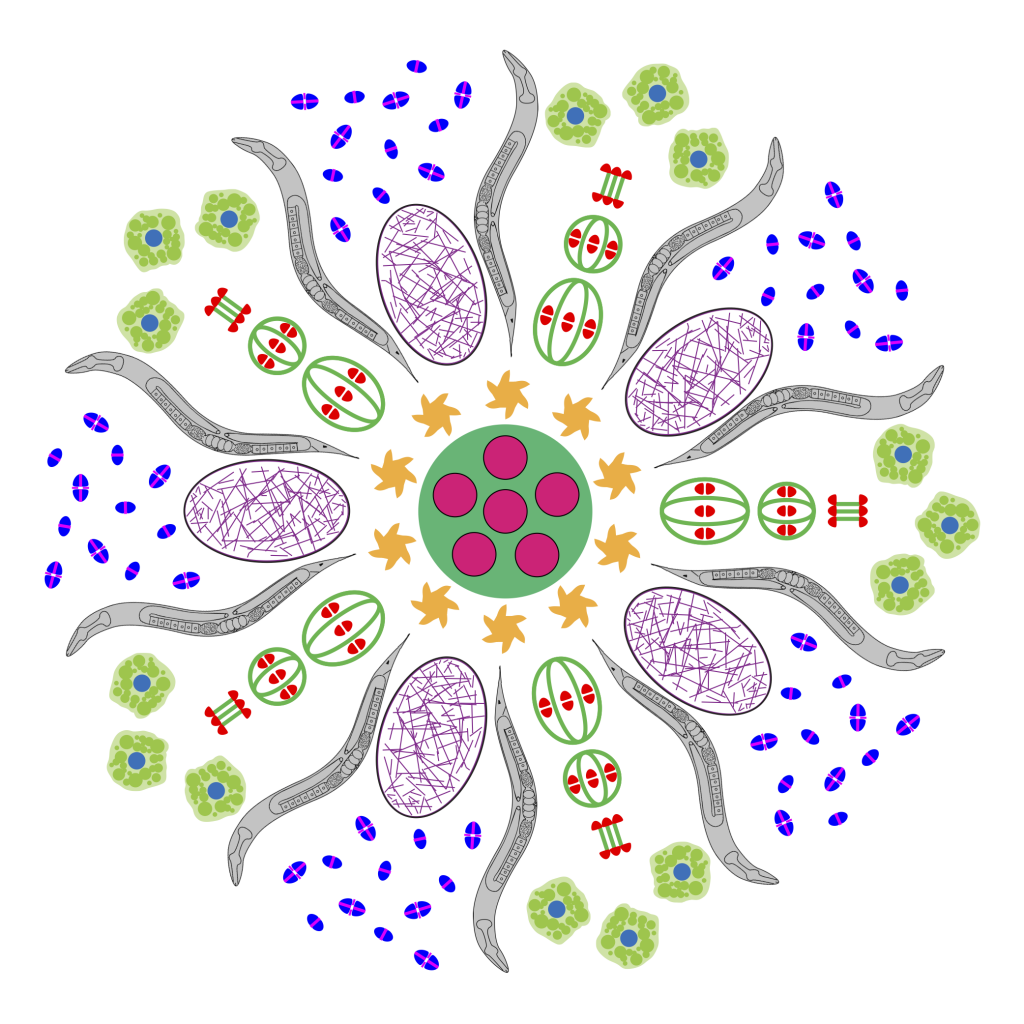
Welcome to the McNally lab. We are located in the Department of Molecular and Cellular Biology at the University of California, Davis. We work on the mechanisms of asymmetric spindle positioning, chromosome segregation, and polar body formation during female meiosis in C. elegans.
Female meiosis is critical for the development of human embryos. Down Syndrome and a high proportion of miscarriages are caused by non-disjunction during female meiosis. In addition, current methods for mammalian cloning involve removing the meiotic spindle from the metaphase II-arrested egg and introducing a G1 diploid nucleus which is converted into a diploid pseudo-meiosis II spindle.
During meiosis in most species, chromosome number is reduced four-fold by first segregating pairs of homologous chromosomes and subsequently segregating pairs of sister chromatids. In male animals, these sequential chromosome segregation events result in four haploid sperm. In contrast, only one of four meiotic products is inherited during female meiosis. This asymmetric inheritance is mediated by meiotic spindles that are attached by one pole to the oocyte cortex at anaphase. Female meiotic spindles in mouse, humans, and C. elegans are considered “anastral” because they do not have visible astral microtubules or the centriole-containing centrosomes that organize astral microtubules. Thus novel mechanisms are likely to mediate meiotic spindle positioning in C. elegans and human oocytes. In fact, mouse meiotic chromosomes translocate to the cortex in the complete absence of spindle microtubules.
Cortical meiotic spindle positioning is followed by asymmetric cell divisions that leave one haploid set of chromosomes in the female pronucleus of a large egg and the “extra” chromosome in tiny cells called polar bodies. There is also conservation in the spindle movements that lead up to the perpendicular cortical attachment of the spindle at anaphase. In mouse, Xenopus, and C. elegans, the meiosis I spindle or its precursor assembles at a distance from the cortex and translocates to the cortex after nuclear envelope breakdown. In C. elegans, Drosophila, Xenopus, and at least meiosis II of mouse, the spindle is initially oriented parallel to the cortex and then undergoes a discrete rotation to the perpendicular orientation. C. elegans, however, is the only genetically-tractable species where the complete sequence of spindle movements during meiosis I and meiosis II can be continuously tracked by time-lapse imaging.
Our lab uses the nematode C. elegans to study the mechanisms of asymmetric meiotic spindle positioning and polar body extrusion. We are specifically interested in understanding how the cytoskeleton contributes to these processes, as well as understanding the molecular mechanisms responsible for driving the spatial and temporal order of events. Some of the current projects in the lab focus on:
– The role of the microtubule motor protein, Kinesin-1, in meiotic spindle positioning.
– The role of the microtubule-severing ATPase, katanin, in meiotic spindle assembly and spindle length control.
– The role of fertilization and the SPE-11 protein in meiotic progression and polar body formation.
– The mechanism of polar body extrusion
– The role and regulation of cytoplasmic dynein dependent meiotic spindle movements.